

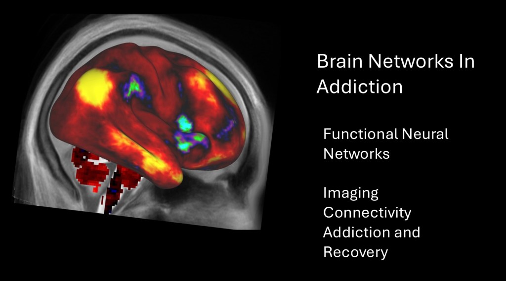
Brain Networks in Addiction
Imaging, Connectivity, Addiction and Recovery
This post explores organization of the brain into large scale networks composed of communicating integrated systems and subsystems. There is a growing body of evidence characterizing the ways in which these systems are altered in substance addictions. This information may then be applied to develop diagnostic and prognostic bio markers along with improved targeted treatments.
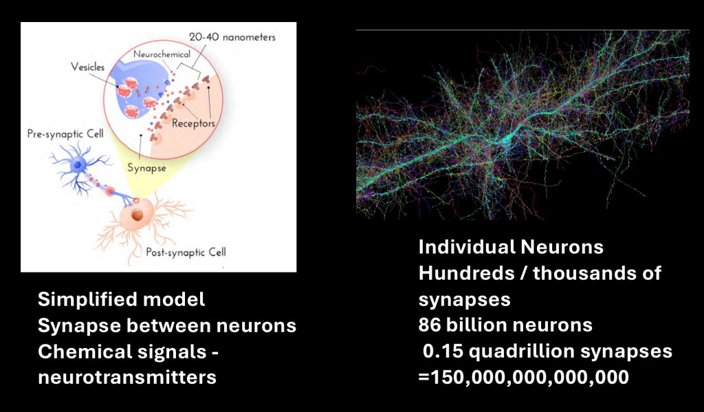
Individual neurons and synapses comprise the smallest functional units in the nervous system. Information is received from dendritic inputs. It is then passed as an electrical impulse down the axon resulting in neurotransmitter release. Communication between neurons is chemical and modulated by various local factors. There are many specialized types and subtypes of neurons found in different brain regions.
This micrograph of an individual neuron illustrates complexity of structure with many thousands of communicating dendrites and synapses. The adult human brain consists of an estimated 86 billion neurons and 0.15 quadrillion synapses.
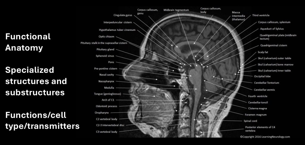
The brain consists of many distinct functional components and sub components. These can be characterized by the role a particular structure plays in functions such as memory, speech, motor function, stress response, and so on. Much of neuroscience is concerned with how each of these components function in normal and pathological states and how they interact with each other in more complex ways.
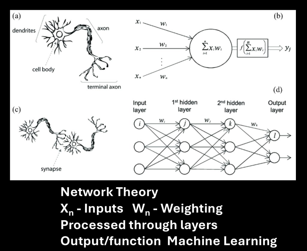
On a larger scale the brain can be thought of in terms of a number of networks integrating multiple spatially distant components into functional domains. Network theory is useful in biology, computer science, social science, communications and other fields.
In the simplified diagram above a single neuron can be represented as consisting of multiple separate inputs (X1, X2,…) each with a separate weighting (W1, W2…). The cell body output can then be represented as a function of the sum of these weighted inputs. These may be excitatory or inhibitory. More of these connected as a network can consist of multiple layers of nodes and connections (edges). This can be represented as a graph as shown above.
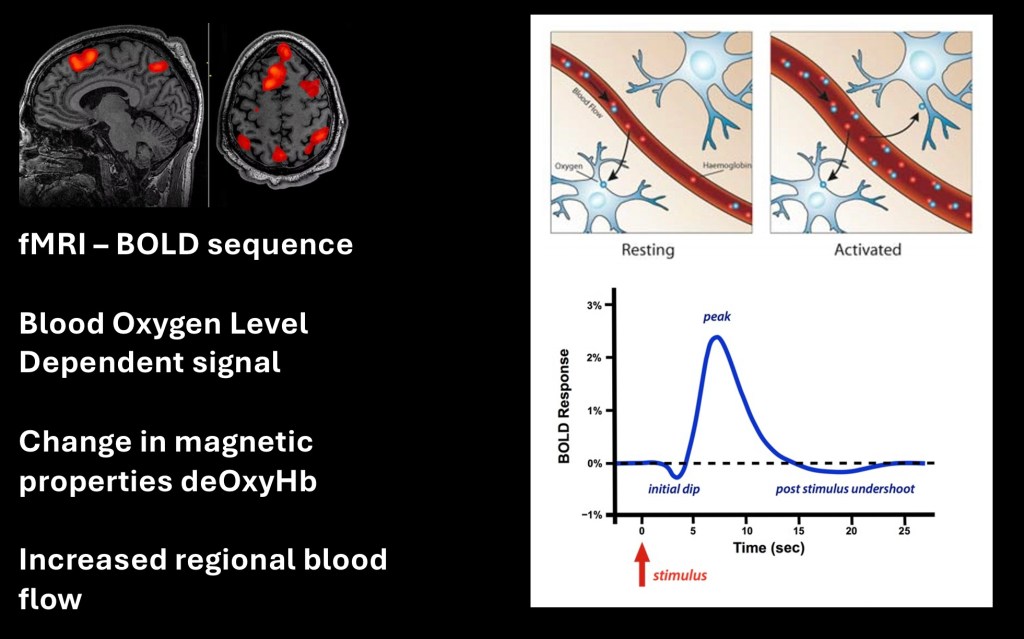
The primary tool used in studies of biological neural networks is functional MRI (fMRI). The name is something of a misnomer as fMRI does not directly measure function. It is an indirect indicator of brain activity.
fMRI is a measure of Blood Oxygen Level Dependent (BOLD) signal. Brain tissue has a very high metabolic demand. When a section of tissue becomes active oxygen demand increases. In response two things occur:
– Oxygen is extracted from hemoglobin in passing blood vessels. When oxyhemoglobin becomes deoxyhemoglobin it alters its magnetic state (to paramagnetic)
– The capillaries carrying blood temporarily dilate increasing blood flow to the active tissue
This results in a very small detectable MR signal change, BOLD signal using a sensitive pulse sequence (echo planar). Keep in mind it is a relative change as the brain is in a constant state of metabolism and activity. It is also transient. Note that the peak occurs several seconds after the stimulus. That is a long time in neural transmission which is measured in milliseconds.
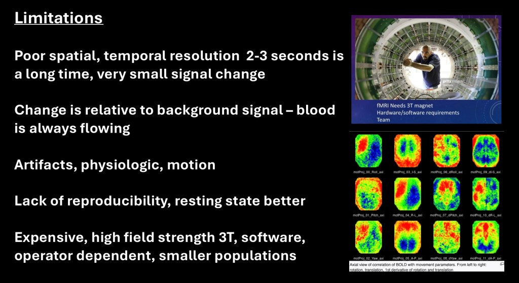
Limitations in fMRI.
Because of the physiologic limitations discussed above and the time required to collect enough signal for imaging, fMRI has poor spatial and temporal resolution. For localization the fMRI data can be superimposed on a standard structural image. Small structures and transient signal changes however, may be lost.
There is a large amount of background signal as well as artifact from motion, heart activity, and breathing. To be useful these have to be extracted and normalized. This adds to the possibility of misinterpretation due to extensive post processing.
Earlier studies focused on task oriented imaging such as having the subject view a picture during acquisition. Many of these studies were found to be non reproducible due to many uncontrolled variables.
MRI is expensive and time consuming. fMRI requires very high field magnets, specialized software, and staffing. It does not lend itself to large population studies and sample sizes are often limited.
Despite these obstacles a large body of research is emerging demonstrating robust reproducible evidence. Resting state MRI (rs fMRI) has been found to be highly consistent across studies. In these studies the subject is instructed to focus on nothing in particular during imaging. Large databases of both normal and abnormal states are now openly accessible and can be used to standardize results.
The advantage is that MRI is noninvasive and an established commercial technology. It provides both structural and functional information in living humans in near real time. As standardized protocols and data have become more available results can be correlated from multiple sources. For addiction studies, as much of the basic science and neuropathology has been worked out, the focus can begin to shift to practical applications, treatment, prevention, and recovery.
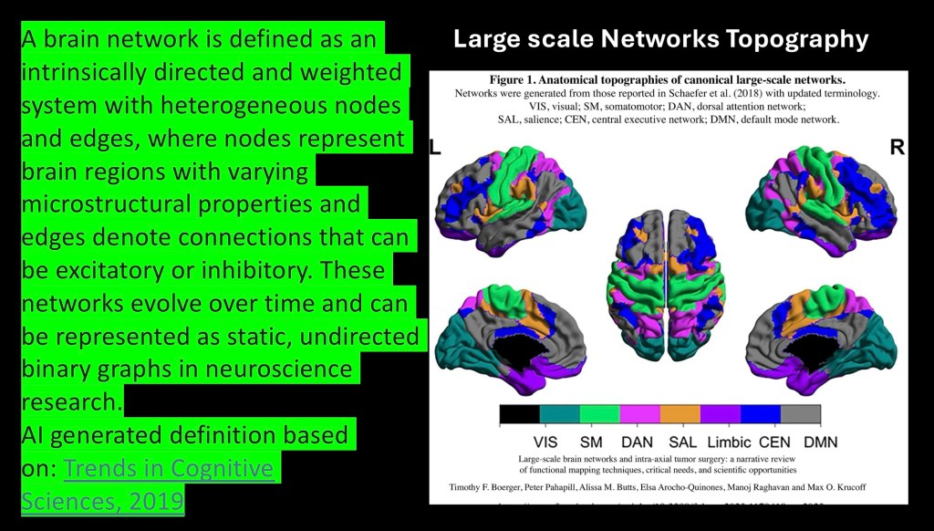
“A brain network can be defined as an intrinsically directed and weighted system with heterogeneous nodes and edges, where nodes represent brain regions with various micro structural properties and edges represent connections that can be excitatory or inhibitory. These networks evolve over time and can be represented as static, undirected binary graphs in neuroscience research.
AI generated definition based on
Tends in Cognitive Sciences 2019
The above color topographical map of the whole brain represents division into seven neural networks. These are generally accepted divisions although there is some variation from different sources.
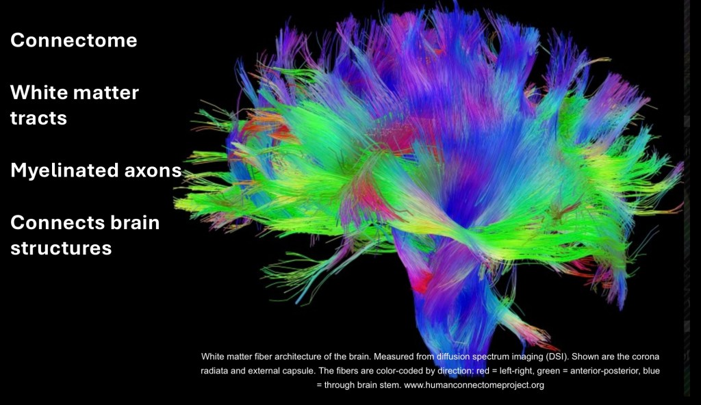
The Connectome consists of the ‘wiring’ between one region of the brain to another. Where the cortex and deep structures (grey matter) consist of synaptic neural tissue the white matter consists of myelinated axons connecting various brain structures. Considerations of dynamic connections both within and between networks is an essential component of analysis.
Large databases such as the human Connectome project are now available as research tools. The above image was constructed with colors coding for direction of fibers: red=left-right, green=anterior-posterior, blue=through brain stem. The images are obtained by MRI diffusion tensor imaging which is sensitive to the movement of water molecules along white matter tracts.
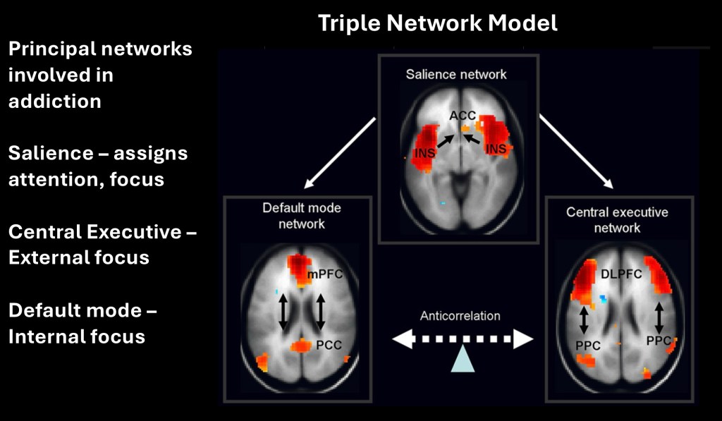
This post will focus on these three networks which have been described in the Triple Network Model. Salience network has the general role of assigning attention and focus to both internal and external states. The Default Mode Network is largely concerned with inward direction and internal focus. The Central Executive Network is more directed toward the external environment and responses. While these are broad generalizations the model serves as a useful framework for further discussion.
Note that Salience serves as a switchboard for the other two networks in this model.
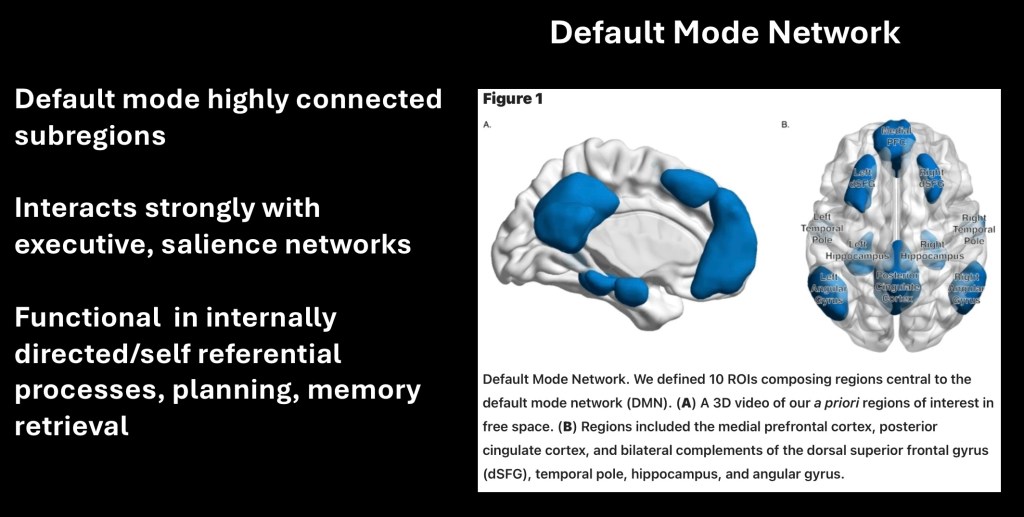
The default node network (DMN) consists of the medial prefrontal cortex, dorsal superior frontal gyrus, posterior cingulate gyrus, temporal pole and hippocampus. The DMN is strongly internally connected and connected to the executive and salience networks.
The DMN is active during periods of inattention however not exclusively. It is primarily active in self referential processes and internal monitoring. Autobiographical and long term memory retrieval and planning are processed through this network.
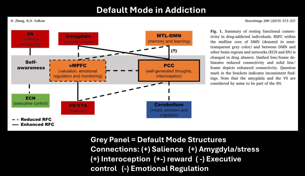
Default Mode Network abnormalities in addiction
The areas in the central gray box represent the central default mode. Solid lines represent increased connectivity and dashed lines decreased connectivity. In addiction Salience input to DMN is increased and Executive is decreased. There is increased connection to the Amygdyla and stress reaction with emotional dysregulation. Focus becomes increasingly internal and self referencing while representative internal monitoring appears to be impaired in addiction.
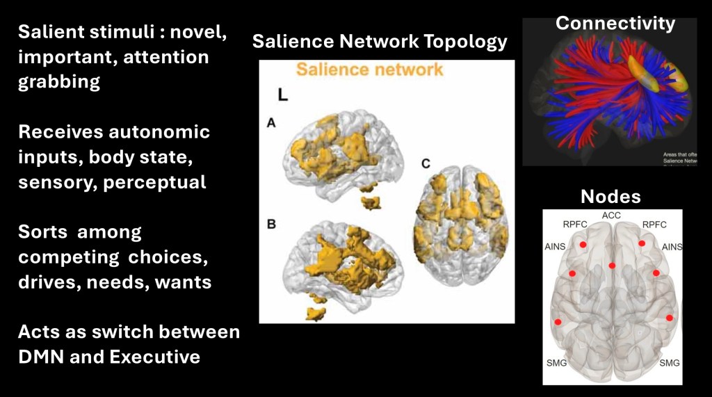
The Salience network primarily consists of the anterior insula within the temporal lobe and the anterior cingulate cortex located deep within the center of the brain. Salience assigns importance and attention to competing stimuli, wants, and needs. It is important to note that the salience network responds to both positive and negative stimuli whereas the reward system primarily responds to positive valence. The network receives both internal and external sensory and perceptual inputs. In addition to substance use disorder imbalance in the network is implicated in other neuropsychiatric disorders including schizophrenia, PTSD, and anxiety disorders.
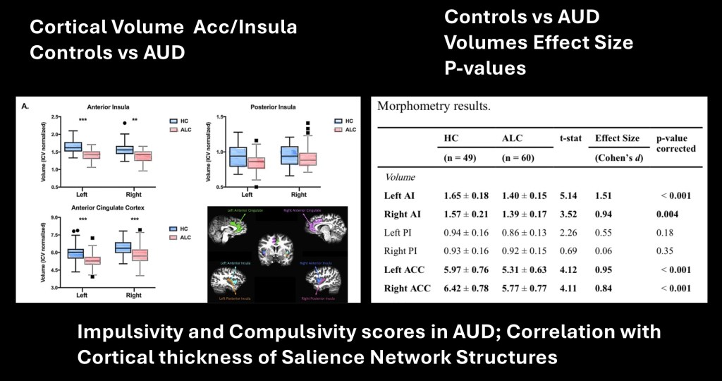
Structural information may be correlated to network and clinical data. There has been an ongoing discussion concerning the importance of global tissue loss due to alcohol toxicity vs focal dysfunctional volume loss. This study looked at the relationship between alcohol use disorder, neuro cognitive markers, and focal cortical volume loss in salience network structures. Voxel based morphometry demonstrated a positive correlation between degree of grey matter volume loss and impulsivity/compulsivity scores on psychometric testing in subjects with Alcohol Use Disorder. Focal volume loss was identified involving the anterior cingulate cortex and anterior insula bilaterally.
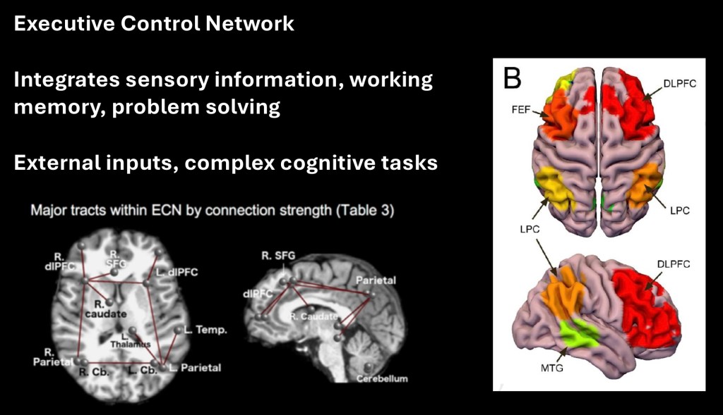
Executive Control Network is composed of the dorsal-lateral prefrontal cortex, lateral parietal lobes, and portions of the temporal lobes bilaterally. Functionally the Executive network is concerned with integrating external sensory information, working memory, and complex cognitive tasks. It governs both initiation of motor activity as well as inhibition of response. Thus it is key in purposeful goal oriented control of complex tasks. The MR images above depict major connectivity tracts bridging ECN centers.
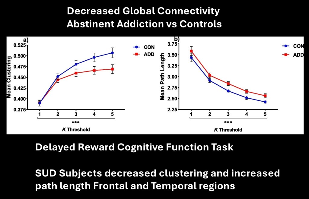
This study looked at abstinent subjects with history of addictions to a number of different substances. In this portion of the study subjects were given a cognitive task, choosing between shapes displayed on a screen, with delayed monetary reward for correct answers. Global connectivity was measured and compared with controls.
The graphs represent clustering of nodes and path lengths across global networks during task performance. These are important in the relative efficiency of information processing within and between networks. Data demonstrate decreased network functional connectivity in abstinent SUD subjects compared with controls.
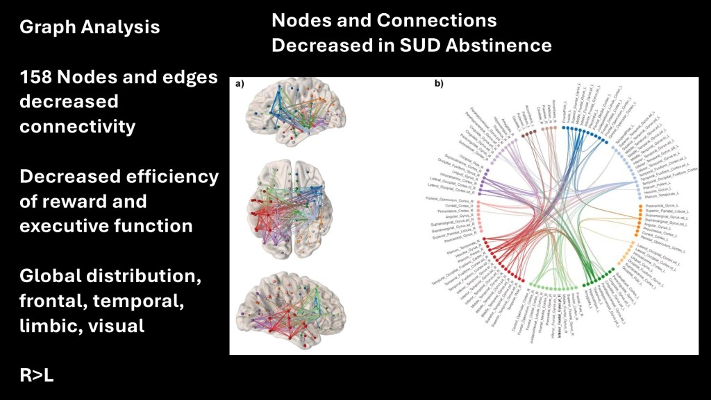
The same study identified 158 separate nodes and edges involving executive function and reward processing with impaired connectivity in subjects with SUD in remission. These occurred in frontal, temporal, limbic and visual systems. More of these were identified in the right hemisphere than in the left.
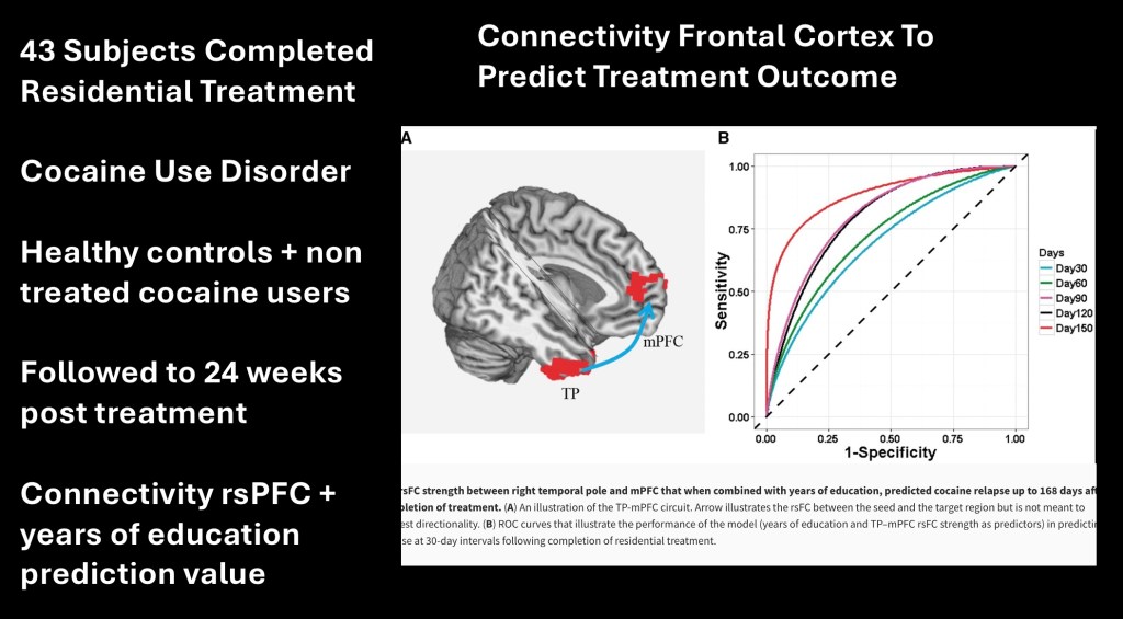
This study looked at outcomes to relapse using rs fMRI in subjects with cocaine addiction following completion of 2-4 weeks of residential treatment. Non-treatment seeking and healthy control subjects were included for comparison. Resting state (rs) fMRI was performed after discharge and to 24 weeks post treatment.
The above receiver operating characteristic curve (ROC) shows sensitivity vs 1-specificity using connectivity between the anterior temporal and medial prefrontal cortex combined with years of education as a predictive marker for abstinence during the study period. The curves toward the upper left are greater in predictive value. The metric demonstrated high predictive value which was greatest at longer time intervals.
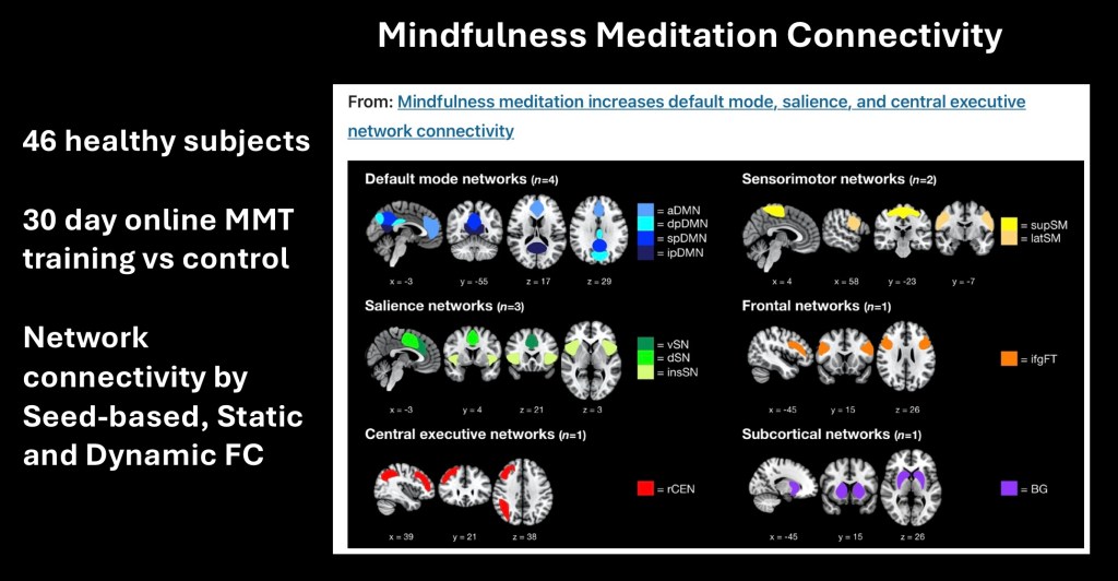
This was a study in healthy subjects (n=46) on effects of mindful meditation (MMT) training in network connectivity. Subjects were assigned to two groups. One group received a 30 day online training course in mindful meditation. The other control group received a general health training course. Connectivity was measured by rs fMRI by seed based, dynamic, and static global connectivity before and after training. The six functional networks measured are pictured above.
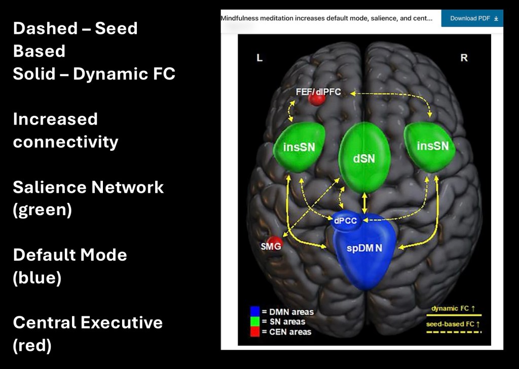
Findings are summarized in this diagram.
Blue = Default Mode
Green = Salience
Red = Executive
Results for both increased seed based (dashed lines) and dynamic (solid lines) connectivity are shown. Overall significantly increased connectivity between default mode network and components of salience and executive networks were identified. As dysfunctional alterations in these areas have been shown in active addiction extending into recovery there is strong evidence for benefit of MMT as part of an overall treatment plan for individuals with this disorder.
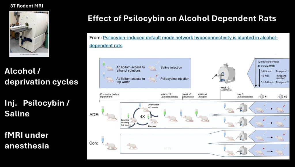
Preclinical animal studies on neural networks can be performed. Suitable equipment and MRI units have been engineered to accommodate small animals. Brain maps and databases exist for rodents.
There has been a resurgence of interest in the effects of psychedelic drugs for treatment of neuropsychiatric disease including addiction. This study looked at effects of psilocybin in an experimental alcohol dependance/relapse paradigm.
A rat strain bred for a proclivity for alcohol was divided into control and alcohol groups. The alcohol test group was given unlimited access to alcohol solution followed by varying cycles of alcohol and deprivation. Under these conditions rats will consume more alcohol on reinstatement. This forms a model for relapse conditions in humans. After a week on just water the rats received either saline or 1mg/kg psilocybin injection and fMRI was performed under anesthesia.
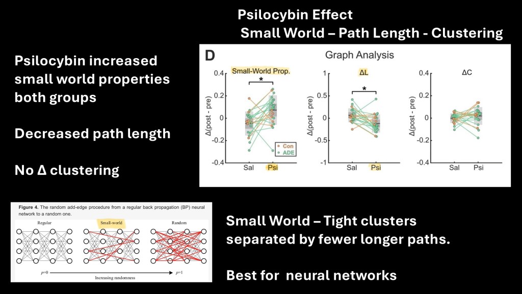
Decreased path length and increased clustering of nodes are measures of small world properties of networks as measured by graph theory. Briefly, certain networks including neural networks more efficiently transfer information when arranged in tight highly connected clusters separated by fewer connections and hubs.
Decreased path lengths and increase in small world connectivity was identified in controls and test subjects following psilocybin administration. This is a drug induced neuroplastic change.
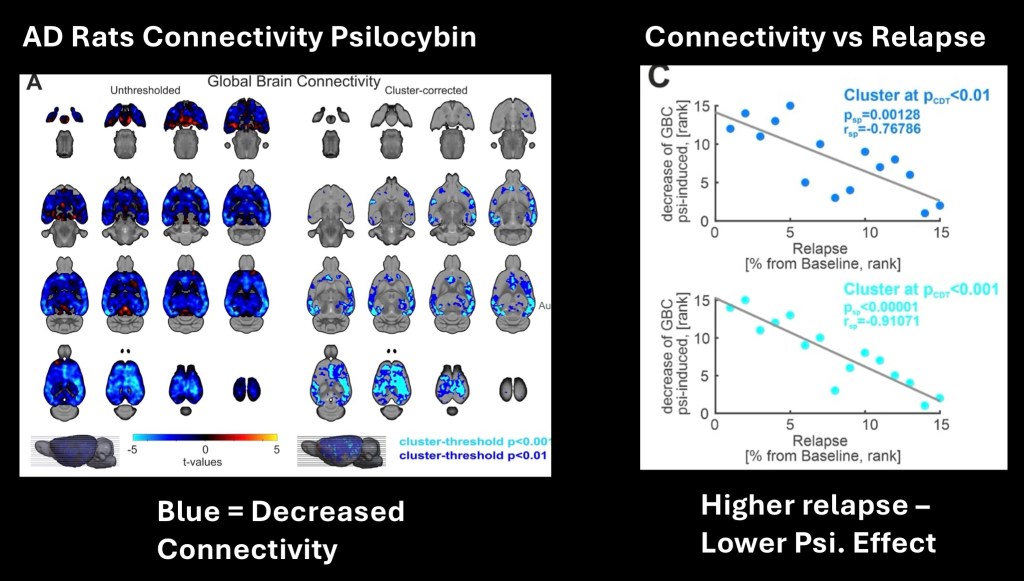
Decreased connectivity resulting in more efficient transmission is shown in blue correcting for clustering of nodes. On the right the psilocybin global brain connectivity (GBC) is shown to be negatively correlated with the relapse rate for individual rats. The more alcohol consumed per relapse cycle, the less observed effect from the standard dose of psilocybin administered. The observed psilocybin neural response was blunted by chronic higher dose alcohol.
This suggests that a therapeutic dose may need to be higher in subjects with active alcohol use disorder to achieve desired effects.
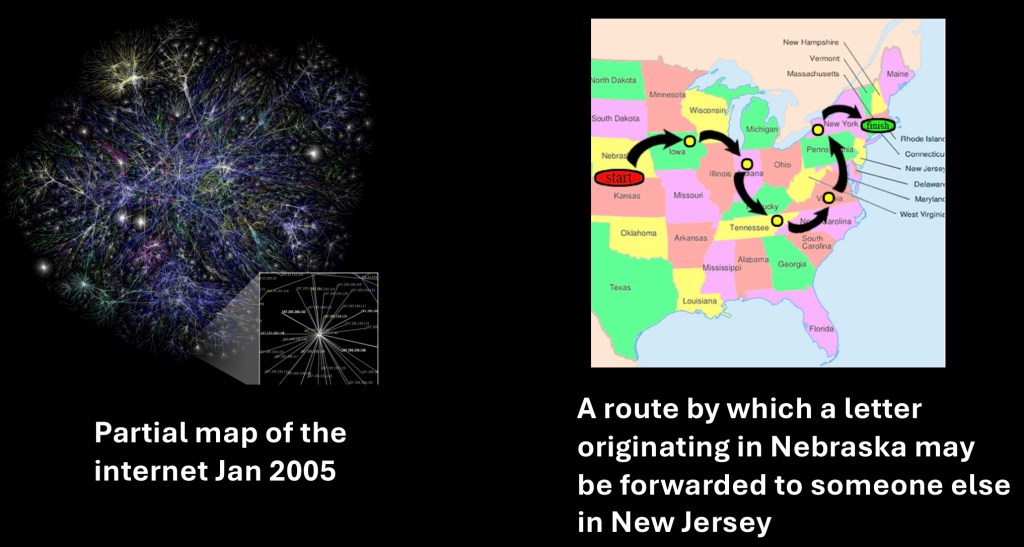
The small world concept originated in experiments and observations on social networks. The idea that “six degrees of separation” separated all individuals on earth has become a popular meme. Many organizations including recovery support groups such as Alcoholics Anonymous or LifeRing follow a small world pattern where relatively small, close knit clusters are connected through hubs to a central organization and mission. The formal concept was published by social scientist Stanley Milgram in the magazine Psychology Today in 1967.
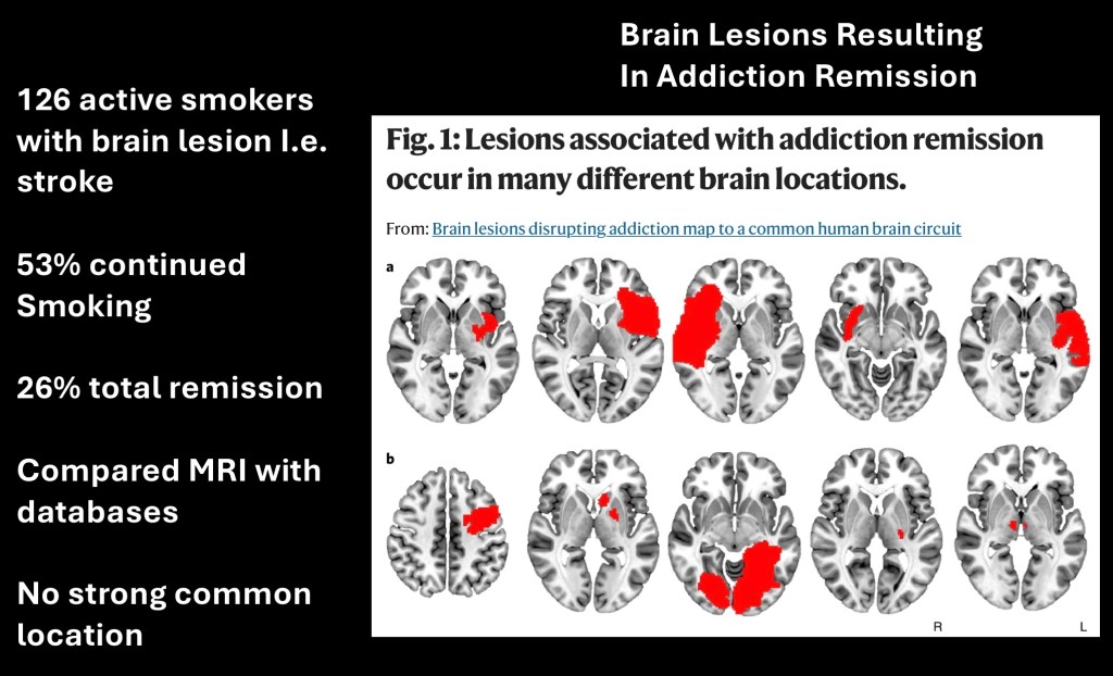
This research study took a different approach. It has been noted that in some individuals with brain lesions such as from a stroke or traumatic brain injury a spontaneous remission of addiction may occur with no cravings or recurrence. This study looked at maps of brain injury in 126 active tobacco smokers 26% of whom had complete remission following the event.
The lesions had no discrete dominant location pattern. The investigators hypothesized that these seemingly unrelated brain injuries may be connected by a common neural pathway which was disrupted by the traumatic event. The investigators correlated the lesions with connectome maps to look for an underlying communication circuit.
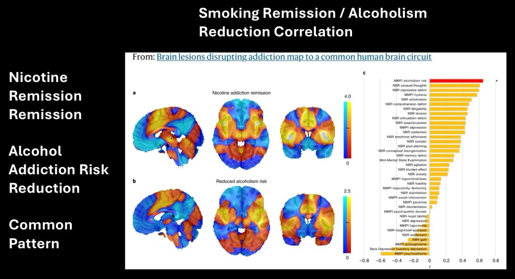
The lesions were correlated with a known tobacco smoking database as well as a general population. The lesions mapped to areas of common positive (warm red-yellow) and negative (cold blue-green) connectivity. The connectome patterns derived from both databases are near identical. Further comparison with a map derived from individuals with alcohol use disorder also yielded nearly the same pattern. The authors suggest a potential common pathway for treatment of substance addictions for further investigation.
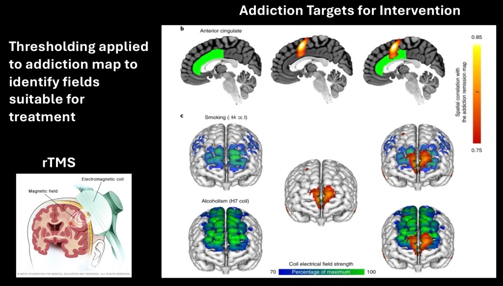
Thresholding was applied to further refine key foci suitable for therapeutic intervention. These were correlated with coil fields which may be used in Transcranial magnetic stimulation. A technique that generates pulses of electromagnetic fields to stimulate brain tissue. A TMS device has recently been approved by the FDA for use as an aid in smoking cessation.
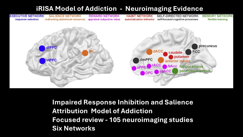
This is from a review article of task based functional MRI studies published in 2018. The article focused on application to the impaired response inhibition and salience attribution model of addiction. This model focuses on deficits in executive function resulting in impaired ability to inhibit drug related responses along with salience network dysfunction.
105 whole brain neuroimaging studies were included in the review. The brain was divided into six functional networks with structures color coded in the above diagram.
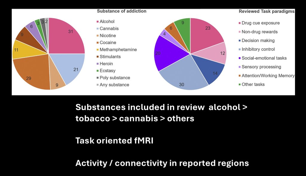
The chart on the left indicates the substances included in the review. The tasks evaluated are represented in the chart on the right.
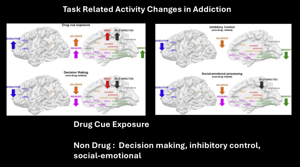
Composite results demonstrated increased activity in all six brain networks included in the studies in SUD subjects when exposed to drug cues. In the iRISA hypothesis these networks are coordinated in forming a response activating attention, memory and executive decision making resulting in drug seeking. Although some differences were noted when engaged in other tasks, delayed and muted responses to non drug related activities were consistently observed.
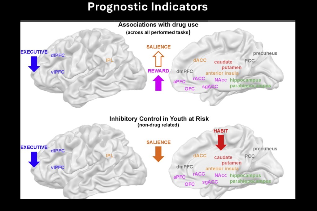
Across all tasks drug dependent subjects demonstrated executive functional impairment. Reward network was hyper active on drug related cues. Salience network was altered from normal controls and inconsistent across different substances. Elevated activation of the reward system was strongly associated with relapse and increased levels of drug use.
Studies on at risk youth noted lower levels of activity in salience and habit forming networks. Adolescents with blunted responses in delayed control tasks had higher incidence of problematic drinking 4 -5 years later.
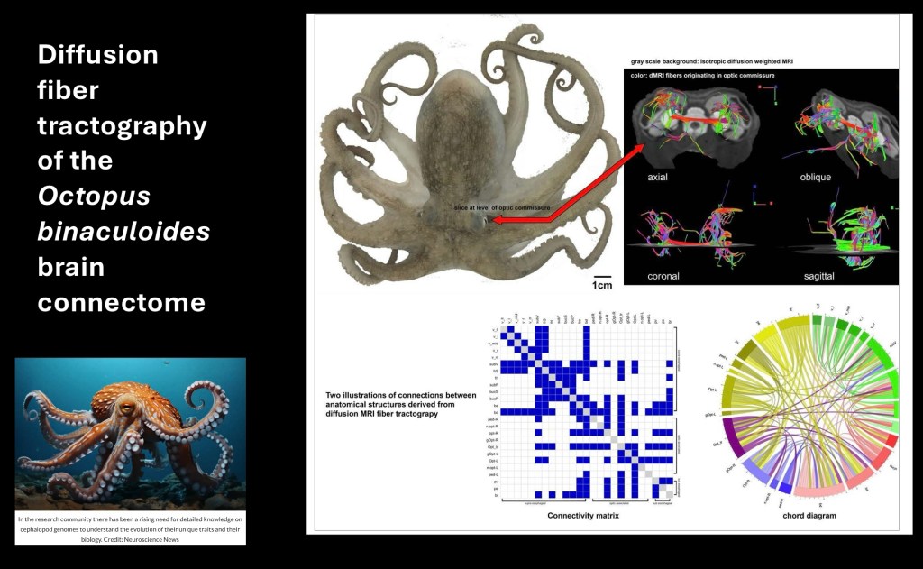
The octopus has a high level of intelligence in mathematical and spatial reasoning. They frequently escape their enclosures. They can use tools and solve puzzles. Structurally however the organization of the octopus nervous system is markedly different from humans and other mammals.
………………………………………………………..,.,,.,……………………
While the concept of the brain acting as a composite of interconnected networks is not a new one it has been a challenge to study in a systematic quantifiable way. Functional MRI provides a platform to study interconnected networks in vivo under controlled conditions. As the neuroscience of addiction has evolved over the past few decades the focus is shifting from basic neurobiological processes to clinical applications, recovery, and prevention.
This post introduced some of the basic principles of fMRI and network theory. Examples of applications involving structural and functional imaging, preclinical laboratory applications, and application to clinical findings and treatment were explored. This is an evolving area of research with significant practical potential.
…………………………………………………………..
JK 8/23

Thank you for your time spent in reviewing this post. Comments and suggestions are always welcome. For information and educational purposes only. Images and data obtained from sources freely available on the World Wide Web. This post should not be considered medical or professional advice.
Jeffk072261@hotmail.com
REFERENCES
Networks
Brain default-mode network dysfunction in addiction
Rui Zhang a, Nora D. Volkow
Brain default m-mode network dysfunction in addiction – ScienceDirect
……………………………………………………….
The effect of seed location on functional connectivity: evidence from an image-based meta-analysis
Front. Neurosci., 30 May 2023
Sec. Brain Imaging Methods
Volume 17 – 2023 | https://doi.org/10.3389/fnins.2023.1120741
Network analysis of substance abuse and dependence
symptoms
Mijke Rhemtullaa, Eiko I. Friedb,*
, Steven H. Aggenc,d Francis Tuerlinckxb Kenneth S.
Kendlerc,d,e, and Denny Borsbooma
Mijke Rhemtulla: MRhemtulla@uva.nl
aDepartment of Psychology, University of Amsterdam, The Netherlands
Volume 185, 15 January t
………………………………………………………
Brain network dysfunctions in addiction: a meta-analysis of
resting-state functional connectivity
Translational Psychiatry (2022)12:41 ; https://doi.org/10.1038/s41398-022-01792-6
………………………………………………………………
Small world connectivity
…………………………………………………………………..
Beyond Functional Localization
Advancing the Understanding of Addiction-Related Processes by Examining Brain Connectivity
Matthew T. Sutherland, Xia Liang, Yihong Yang, and Elliot A. Stein
The Wiley Handbook on the Cognitive Neuroscience of Addiction, First Edition. Edited by Stephen J. Wilson.
© 2015 John Wiley & Sons, Ltd. Published 2015 by John Wiley & Sons, Ltd.
………………………………………………………………………….
Connecting Circuits with Networks in Addiction Neuroscience: A Salience Network Perspective
by Adriana K. Cushnie 1, Wei Tang
Int. J. Mol. Sci. 2023, 24(10), 9083; https://doi.org/10.3390/ijms24109083
……………………………………………………………………….
……………………………………………………………………….
………………………………………………………………………
Triple Network Resting State Connectivity Predicts Distress Tolerance and Is Associated with Cocaine Use
Elizabeth D. Reese 1, Jennifer Y. Yi 1, Katlyn G. McKay 1 , Elliot A. Stein 2, Thomas J. Ross 2 and Stacey B. Daughters
J. Clin. Med. 2019, 8, 2135; doi:10.3390/jcm8122135
………………………………………………………….
Exploring the brain network: A review on resting-state fMRI functional connectivity
Martijn P. van den Heuvel ⁎, Hilleke E. Hulshoff Pol
Rudolf Magnus Institute of Neuroscience, University Medical Center Utrecht, Neuroimaging Division, The Netherlands
European Neuropsychopharmacology (2010) 20, 519–534
………………………………………………….
Small-World Brain Networks Revisited
Danielle S. Bassett1,2 and Edward T. Bullmore3,4
The Neuroscientist
2017, Vol. 23(5) 499–516
© The Author(s) 2016
Reprints and permissions: sagepub.com/journalsPermissions. https://www.ncbi.nlm.nih.gov/pmc/articles/PMC5603984/pdf/10.1177_1073858416667720.pdf
…………………………………………………….
BOLD imaging | Radiology Reference Article | Radiopaedia.org
Addiction Related Alteration in Resting-state Brain Connectivity Ning Maa, Ying Liub, Nan Lia, Chang-Xin Wangb,
. Neuroimage. 2010 January 1; 49(1): 738–744. doi:10.1016/j.neuroimage.2009.08.037.
………………………………………………………
Interactions between the Salience and Default-Mode Networks Are Disrupted in Cocaine Addiction
Xia Liang, Yong He, Betty Jo Salmeron, Hong Gu, Elliot A. Stein and Yihong Yang
Journal of Neuroscience 27 May 2015, 35 (21) 8081-8090; https://doi.org/10.1523/JNEUROSCI.3188-14.2015
…………………………………………………………
Salienceaddiction
……………………………………………………………..
………………………………………………………………
fMRI BOLD signal
………………………………………………………………..
Abnormal Brain Default-Mode Network Functional Connectivity in Drug Addicts
- Ning Ma ,Ying Liu Xian-Ming Nan Li, Published: January 26, 2011
- https://doi.org/10.1371/journal.pone.001656 PLOS
Abnormal Brain Default-Mode Network Functional Connectivity in Drug Addicts | PLOS ONE
Impaired Functional Connectivity Within and Between Frontostriatal Circuits and Its Association With Compulsive Drug Use and Trait Impulsivity in Cocaine Addiction
Yuzheng Hu, PhD1; Betty Jo Salmeron, MD1; Hong Gu, PhD1; et al
Elliot A. Stein, PhD1; Yihong Yang, PhD1
Author Affiliations Article Information
JAMA Psychiatry. 2015;72(6):584-592. doi:10.1001/jamapsychiatry.2015.1
………………………………………………………………………
Cortico-Striatal-Thalamic Loop Circuits of the Orbitofrontal Cortex: Promising Therapeutic Targets in Psychiatric Illness
Front. Syst. Neurosci., 26 April 2017
Volume 11 – 2017 | https://doi.org/10.3389/fnsys.2017.00025
Effects of Methylphenidate on Resting-State Functional Connectivity of the Mesocorticolimbic Dopamine Pathways in Cocaine Addiction
Anna B. Konova, MA1,2; Scott J. Moeller, PhD1; Dardo Tomasi, PhD3; et al
JAMA Psychiatry. 2013;70(8):857-868. doi:10.1001/jamapsychiatry.2013.1129
https://jamanetwork.com/journals/jamapsychiatry/fullarticle/1699378
………………………………………………………………..
Neuroimaging markers of glutamatergic and GABAergic
systems in drug addiction: relationships to resting-state
functional connectivity
Scott J. Moeller1
, Edythe D. London1,2, and Georg Northoff3
1Departments of Psychiatry and Neuroscience, Icahn School of Medicine at Mount Sinai
Neurosci Biobehav Rev. 2016 February ; 61: 35–52. doi:10.1016/j.neubiorev.2015.11.010.
Joutsa, J., Moussawi, K., Siddiqi, S.H. et al. Brain lesions disrupting addiction map to a common human brain circuit. Nat Med 28, 1249–1255 (2022). https://doi.org/10.1038/s41591-022-01834-y
https://www.nature.com/articles/s41591-022-01834-y
……………………………………………………………
Brain connectivity: an opening window into addiction
Vince D Calhoun1,2
1The Mind Research Network, Albuquerque, New Mexico 87131
2Dept. of ECE, University of New Mexico, Albuquerque, New Mexico
……………………………………………………………………
Shared network-level functional alterations across substance use disorders: A multi-level kernel density meta-analysis of resting-state functional connectivity studies
Arezoo Taebi1 | Benjamin Becker
Addiction Biology. 2022;27:e13200. wileyonlinelibrary.com/journal/adb 1 of 10 https://doi.org/10.1111/adb.13200
https://onlinelibrary.wiley.com/doi/pdfdirect/10.1111/adb.13200
Human Connectome Project | Mapping the human brain connectivity
………………………………………………………………………….
Psilocybin-induced default mode network hypoconnectivity is blunted in alcohol-dependent rats
Jonathan R. Reinwald, Christian N. Schmitz, Ivan Skorodumov
volume 13, Article number: 392 (2023
Brain network dysfunctions in addiction: a meta-analysis of resting-state functional connectivity
Serenella Tolomeo & Rongjun Yu
volume 12, Article number: 41 (2022
……………………………………………………………………
Disturbances across whole brain networks during reward anticipation in an abstinent addiction population
Author links open overlay panel
Liam J. Nestor a John Suckling Karen D. Ersche Anna Murphy c
…………………………………………………………………………..
https://www.sciencedirect.com/topics/neuroscience/brain-network
………………………………………………………………………..
Salience and default mode network dysregulation in chronic cocaine users predict treatment outcome
Xiujuan Geng, Yuzheng Hu, Hong Gu, Betty Jo Salmeron, Bryon Adinoff,
Brain, Volume 140, Issue 5, May 2017, Pages 1513–1524, https://doi.org/10.1093/brain/awx036
………………………………………………………………………..
Salience network structural integrity predicts executive impairment in alcohol use disorders
Caterina Galandra, Gianpaolo Basso, Marina Manera, Chiara Crespi, Ines Giorgi, Giovanni Vittadini, Paolo Poggi & Nicola Canessa
Scientific Reports volume 8, Article number: 14481 (2018)
Mindfulness meditation increases default mode, salience, and central executive network connectivity
Benno Bremer, Qiong Wu, María Guadalupe Mora Álvarez, Britta Karen,
…………………………………………………………………..
RestingState_review.pdf – Google Drive
Resting State functional loci for addiction medicine
……………………………………………..
Small-world network – Scholarpedia
………………………………………………..
The Gray Matter Structural Connectome and its Relationship to
Alcohol Relapse – Reconnecting for Recovery
Susanne G. Mueller., Dieter J. Meyerhoff1
1.Center for Imaging of Neurodegenerative Diseases, Dept. of Radiology and Biomedical Imaging,
University of California, San Francisco, CA. USA
Addict Biol . 2021 January ; 26(1): e12860. doi:10.1111/adb.12860.
Neuroimaging Impaired Response Inhibition and Salience Attribution in Human Drug Addiction: A Systematic Review
, 6 June 2018, Pages 886-903
Author links open overlay panel
…………………………………………………………….
Structural and Functional Brain Recovery in Individuals with Substance Use Disorders During Abstinence: A Review of Longitudinal Neuroimaging Studies
Muhammad A Parvaz, PhD1,2*‡, Rachel A. Rabin, PhD3*, Faith Adams1,2 & Rita Z. Goldstein1,2, PhD
1Department of Pyschiatry, Icahn School of Medicine at Mount Sinai, New York, NY 10029 2Department of Neuroscience, Icahn School of Medicine at Mount Sinai, New York, NY 10029 3Department of Psychiatry, McGill University
………………………………………………………….
Does an Over-Connected Visual Cortex Undermine Efforts to Stay Sober After Treatment for Alcohol Use Disorder?
Angela M. Muller* and Dieter J. Meyerhoff
Department of Radiology and Biomedical Imaging, University of California, San Francisco, San Francisco, CA, United States
ORIGINAL RESEARCH
published: 10 December 2020 doi: 10.3389/fpsyt.2020.536706
…………………………………………………………..
Diffusion MRI Connections in the Octopus Brain
……………………………………………………………
Structural deficits in salience network regions are associated
with increased impulsivity and compulsivity in alcohol
dependence
Erica N. Grodina,Carlos R. Cortesa Primavera A. Spagnoloc, Reza Momenana,*
aClinical Neuroimaging Research Core, NIAAA, NIH, Bethesda, MD 20892, United States
Drug Alcohol Depend . 2017 October 01; 179: 100–108. doi:10.1016/j.drugalcdep.2017.06.014.
…………………………………………………………
Targeting the Salience Network: A Mini-Review on a Novel Neuromodulation Approach for Treating Alcohol Use Disorder
Front. Psychiatry, 16 May 2022
Sec. Addictive Disorders
Volume 13 – 2022 | https://doi.org/10.3389/fpsyt.2022.893833
The relationship between executive functioning and addictive behavior: new insights from a longitudinal community study
Original Investigatio Open access Published: 03 October 2022 Volume 239, pages 3507–3524, (2022) Cite this article
, Kräplin, A., Joshanloo, M., Wolff, M. et al.
https://link.springer.com/article/10.1007/s00213-022-06224-3#citeas
………………………………………………………………………………………………………………
Small-world experiment – Wikipedia
……………………………………………………………….
Why the Octopus Brain is so Extraordinary | Smithsonian Ocean
……………………………………………………………
Disrupted Ventromedial Prefrontal Function, Alcohol Craving, and Subsequent Relapse Risk
Dongju Seo, PhD, Cheryl M. Lacadie, BA, Keri Tuit, PsyD, Kwang-Ik Hong, MS, R. Todd Constable, PhD, and Rajita Sinha, PhD
Departments of Psychiatry (Drs Seo, Tuit, and Sinha, and Mr Hong) and Diagnostic Radiology (Ms Lacadie and Dr Constable), Yale University School of Medicine, New Haven, Connecticut
JAMA Psychiatry. 2013 July ; 70(7): 727–739. doi:10.1001/jamapsychiatry.2013.762.
…………………………………………………………..
Neuroimaging Impaired Response Inhibition and Salience Attribution in Human Drug Addiction: A Systematic Reviewj
Anna Zilverstand, Anna S. Huang, Nelly Alia-Klein Rita Z. Goldstein 1 2k
Neuron Volume 98, Issue 5 6 June 2018, Pages 886-903

Leave a comment