

A brief history of addiction neuroscience
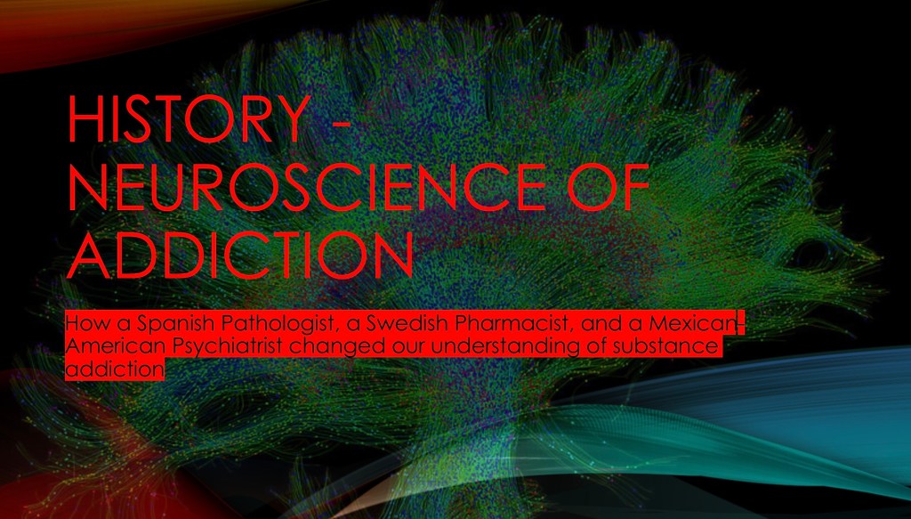
A SHORT HISTORY OF
ADDICTION NEUROSCIENCE
The idea that there is, or should be, a scientific basis for addiction is a relatively new concept. Addiction neuroscience is now well established and its origins can be traced back to the early foundations of our understanding of the structure and function of the central nervous system.
Addiction, or Substance Use Disorder as it is now named, is a robust area of neurobiology.
There is a story here, to tell that story this presentation focuses on the work of three people who have been at key turning points in the interface of science and substance addiction.
As a young man Ramon Cajal had wanted to become an artist. His father was having none of that and off he went to Barcelona to study medicine. He ended up remaining there as professor of anatomy and pathology focusing on the human brain.
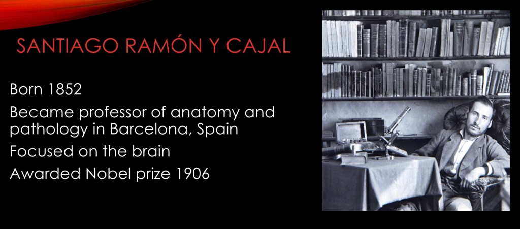
Here he is seated with his microscope, sketch pad, and pen where he was to spend most of his life drawing what he was seeing.
Most of his work was done around the late 1880s. At that time there were advances in slide preparations and tissue staining which improved microscopic observation.
Major advances were made in the study of anatomy and tissue structure in that era.

Cajal made three key observations which form the basis of neuroscience to this day.
He stated that neurons were not directly connected. They were individual cells separated by a space, the synapse.
Cajal observed that neurons were not fixed, they gained and lost synapses over time. He proposed that this was the basis of learning. We call this neuroplasticity today.
He said that unlike other tissues, the nervous system cannot regenerate. It cannot grow new cells.
Using his artistic talents and new tissue staining and preparation techniques he was able to see the structure of individual brain cells in great detail. Until that time it was thought that brain cells were directly connected, transmitting electrical signals throughout the network. His diagrams, like the cerebellar neuron shown here, are still in use today. Note the labeled cell body, axon, and many dendrites. There are about 1,000 trillion synapses in the human brain.
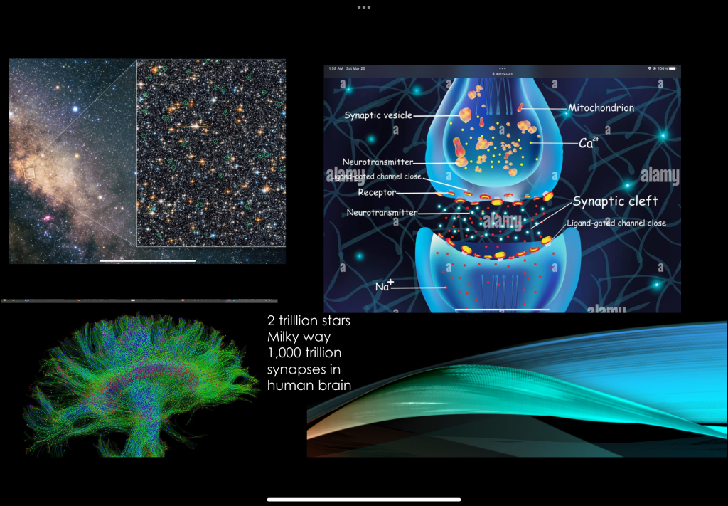
THE SYNAPSE

While much was known about the structure of the brain and other organs it took some time to find what was happening at the biochemical level in cellular function.
Biochemistry and cell biology were to become the next frontier.

Not exactly a household name, Arvid Carlsson was a Swedish professor of pharmacology. He was most active in the 1950s through 1980s. This was an era of great advances in biochemistry and cell biology.
He had been looking at the effects of the drug reserpine which produced temporary paralysis in lab animals. His hypothesis was that a neurotransmitter was involved. Neurotransmitters were a new concept and poorly understood. Early efforts with norepinephrine to reverse paralysis did not work so they tried l-dopa, the precursor of dopamine. While dopamine had been isolated it’s function in the brain was previously unknown.
L-dopa, the immediate precursor to dopamine reversed the paralysis. His work established the role of dopamine as a neurotransmitter and began to localize it to specific regions of the brain. He was eventually awarded a Nobel prize for his work in neurotransmitters.

This led to further investigation into the function of dopamine in other brain functions. Animal experiments found a dopamine spike in the nucleus accumbens following amphetamine administration. Similar findings were seen with other addictive drugs leading to discovery of the reward pathway. This pathway was also shown to be activated in response to other rewarding stimuli such as food and sex.

The central components of the reward pathway consist of the Ventral Tegmental Area in the midbrain extending to the nucleus accumbens. From there the pathway extends to the frontal cortex.
NEUROIMAGING
Through the 1980s to today advances in non invasive imaging technology have opened paths to study the neural basis of addiction in ways not possible previously. Magnetic Resonance Imaging (MRI) and Positron EmissionTomography (PET) allow for characterization of structural and cellular changes under safe controlled conditions in humans.
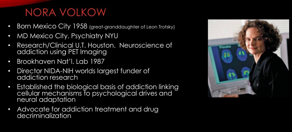
Nora Volkow has been director of the National Institute on Drug Abuse since 2003. She was convinced early in her career that addiction was not a moral failing or character flaw. Addiction has been shown to be a pathological process in brain structure and function which could be proven by application of scientific methods using the tools of neuroimaging. Short and long term changes in neural function and structure have since been demonstrated and reproduced in thousands of published studies.
The NIDA, a branch of the NIH is by far the largest funder of addiction research in the world.

PET study demonstrating changes in dopamine transporter concentration in control subjects, long term abstinent heroin users, and methadone maintenance treated individuals. The transporter protein regulates dopamine concentration in the neural synapse.
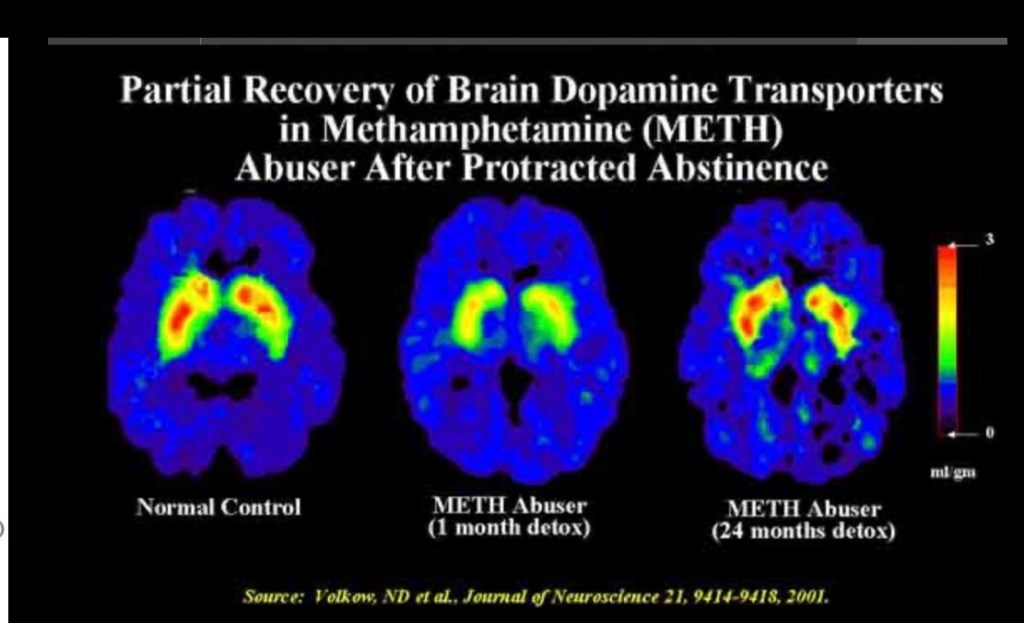
Both short and long term effects have been demonstrated in dopaminergic neurons with partial recovery after long term abstinence. Availability of dopamine D2 receptors have been positively associated with drug craving and risk of relapse. This occurs in alcohol, cocaine, amphetamine, as well as opiate addictions.

Activity reflects glucose metabolism. Diminished frontal lobe metabolism is shown in cocaine addiction. Frontal cortical changes have been associated with impulsive behavior, risk of relapse, and drug craving in human and animal models. The frontal cortex is responsible for executive function, decision making, and cognition. It activates limbic system structures involved in stress reaction, emotion, and memory.
Thank you for your time and consideration in reading this brief introduction. Additional posts will look at neurobiological findings in more detail. As always any feedback is welcome.
For educational and informational purposes only. No commercial or institutional affiliation. This does not constitute medical advice.
REFERENCES
Ehrlich, Benjamin; The Father of Modern Neuroscience Discovered the Basic Unit of the Neurosystem, Scientific American, April 1,2022;
……………………
Yeragani, Vikram et. al. Indian J Psychiatry. 2010 Jan-Mar; 52(1): 87–88
PMCID: PMC2824994
PMID: 20174530
Arvid Carlsson and the story of dopamine
https://www.ncbi.nlm.nih.gov/pmc/articles/PMC2824994/
………………………
J Neurol Neurosurg Psychiatry 2000;68:685–690 685 EDITORIAL
Dopamine agonists: their role in the treatment of Parkinson’s disease
………………………
Cognitive Control of Drug Craving Inhibits Brain Reward Regions in Cocaine Abusers
Nora D. Volkow, M.D.,1,2,* Joanna S. Fowler, Ph.D.,3 Gene-Jack Wang, M.D.,3 Frank Telang, M.D.,2 Jean Logan, Ph.D.,3 Millard Jayne, R.N.,2Yeming Ma, Ph.D.,2 Kith Pradhan, M.S.,3Christopher Wong, M.S.,3
Neuroimage. 2010 Feb 1; 49(3): 2536.
Published online 2009 Nov 11. Doi
https://www.ncbi.nlm.nih.gov/pmc/articles/PMC2818484/
…………………….
Addiction: Beyond dopamine reward circuitry
PNAS
Nora D. Volkow nvolkow@nida.nih.gov, Gene-Jack Wang, Joanna S. Fowler, +1 , and Frank Telang
Edited by Donald W. Pfaff, The Rockefeller University, New York, NY, and approved November 9, 2010 (received for review August 31, 2010)
March 14, 2011
https://www.pnas.org/doi/10.1073/pnas.1010654108
……………………

Leave a comment